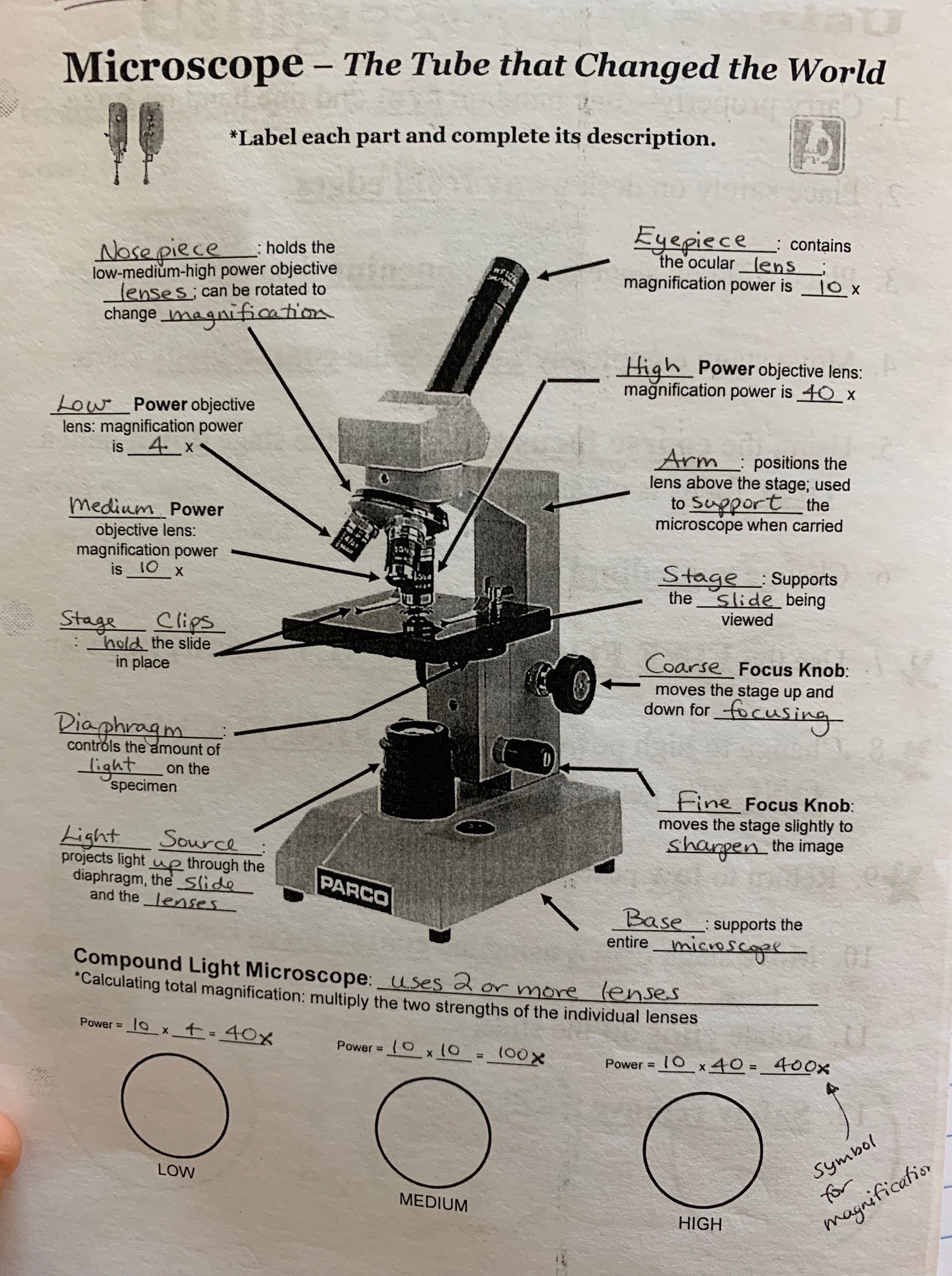
38 microscope with labels and functions
LAS X Industry Microscope software for Industry | Products Microscope software platform for industrial applications to deliver reliable results with confidence in quality control and materials research. ... Activate all relevant functions (e.g. for illumination settings, camera ... Add labels for easy analysis. Apply measurements to several images to determine statistical trend and compare data in ... Microscope Parts, Function, & Labeled Diagram - slidingmotion Microscope parts labeled diagram gives us all the information about its parts and their position in the microscope. Microscope Parts Labeled Diagram The principle of the Microscope gives you an exact reason to use it. It works on the 3 principles. Magnification Resolving Power Numerical Aperture. Parts of Microscope Head Base Arm Eyepiece Lens
Simple Microscope - Parts, Functions, Diagram and Labelling Parts of the optical parts are as follows: Mirror - A simple microscope has a plano-convex mirror and its primary function is to focus the surrounding light on the object being examined. Lens - The biconvex lens is placed above the stage and its function is to magnify the size of the object being examined.

Microscope with labels and functions
Label the microscope — Science Learning Hub 08.06.2018 · All microscopes share features in common. In this interactive, you can label the different parts of a microscope. Use this with the Microscope parts activity to help students identify and label the main parts of a microscope and then describe their functions.. Drag and drop the text labels onto the microscope diagram. If you want to redo an answer, click on the … Parts of a Compound Microscope and Their Functions - NotesHippo Compound microscope uses in forensic labs it easy to detect human fingerprints. A compound microscope can be used to detect the presence of metals. The use of a compound microscope makes studying germs and viruses much easier. Compound microscope uses in schools makes learning biology easy for all children. A FINE GEORGE II MAHOGANY CASED CUFF PATTERN MONOCULAR MICROSCOPE A FINE GEORGE II MAHOGANY CASED CUFF PATTERN MONOCULAR MICROSCOPEJOHN CUFF, LONDON, MID 18th CENTURYThe body tube with stepped moulded shuttered eyepiece over ogee waist and objective tube incorporating marks for six positions on an exponential scale numbered 1 to 6, supported via a tapered collar set in a ring attached to a vertical slide moving …
Microscope with labels and functions. Histology at SIU, tissue prep 16.06.2022 · Trichrome stain. Trichrome uses three dyes (hence the name), including one that is specific for the extracellular protein collagen.Depending on the particular stain combination, a trichrome stain may color collagen fibers sky-blue or bright green. The principle use for trichrome is to differentiate collagen from other eosinophilic structures, such as muscle fibers. Compound Microscope Parts - Labeled Diagram and their Functions Two adjustment knobs are used to focus the microscope: fine focus knob and coarse focus knob. Both knobs can move the stage up and down. You should use the coarse focus knob to bring the specimen into approximate or near focus. Then you use the fine focus knob to sharpen the focus quality of the image. A Study of the Microscope and its Functions With a Labeled Diagram ... A Study of the Microscope and its Functions With a Labeled Diagram To better understand the structure and function of a microscope, we need to take a look at the labeled microscope diagrams of the compound and electron microscope. These diagrams clearly explain the functioning of the microscopes along with their respective parts. LAS X Industry Microscope software for Industry | Products ... Measure parameters, such as the length, area, diameter, angle, or perimeter of objects you mark with adjustable tracing lines, drawing directly in the live images. Add labels for easy analysis. Apply measurements to several images to determine statistical trend and compare data in measurement templates.
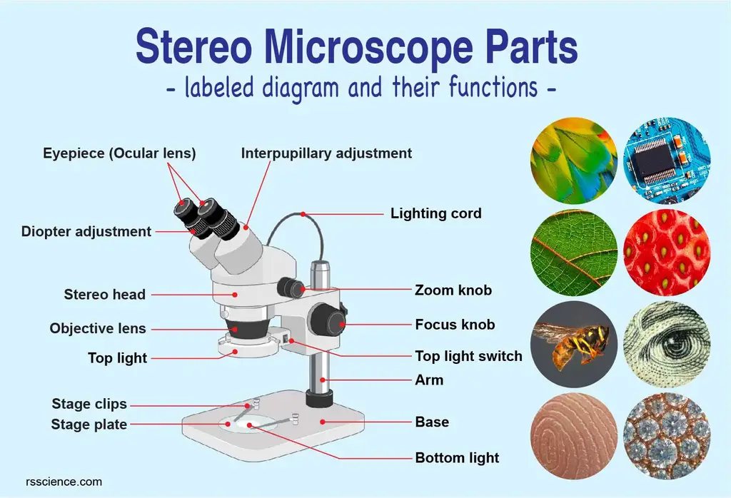
Parts of Stereo Microscope (Dissecting microscope) – labeled … Unlike a compound microscope that offers a flat image, stereo microscopes give the viewer a 3-dimensional image that you can see the texture of a larger specimen. [In this image] Examples of Stereo & Dissecting microscopes. Major microscope brands (Zeiss, Olympus, Nikon, Amscope, Omano, Leica …) all produce stereomicroscopes. Microscopy- History, Classification, Terms, Diagram - The Biology Notes History of Microscope. In the 1 st Century AD, the Romans invented the glass and used them to magnify objects. In the early 14 th Century AD, eyeglasses were made by Italian spectacle makers. In 1590, two Dutch spectacle makers, Hans, and Zacharias Jansen created the first microscope. It was a simple tube with 2 lenses system and had 9X ... Parts of a microscope with functions and labeled diagram - Microbe Notes Microscopes are instruments that are used in science laboratories to visualize very minute objects such as cells, and microorganisms, giving a contrasting image that is magnified. Microscopes are made up of lenses for magnification, each with its own magnification powers. Compound Microscope Parts, Functions, and Labeled Diagram Compound Microscope Parts, Functions, and Labeled Diagram Parts of a Compound Microscope Each part of the compound microscope serves its own unique function, with each being important to the function of the scope as a whole.
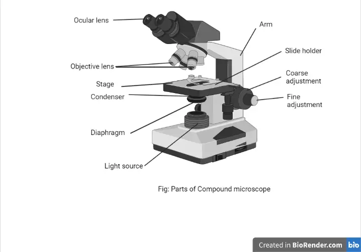
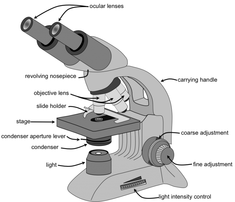
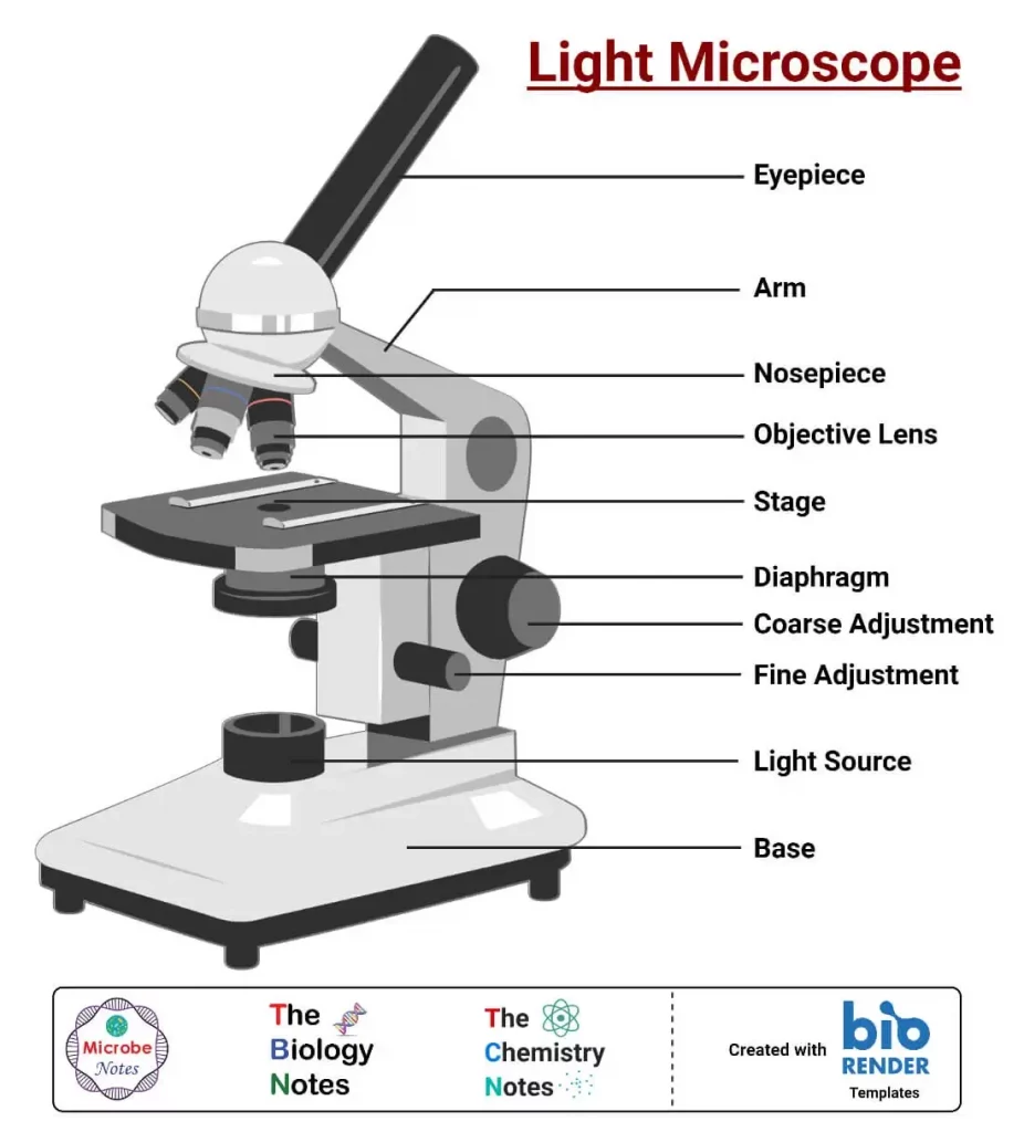
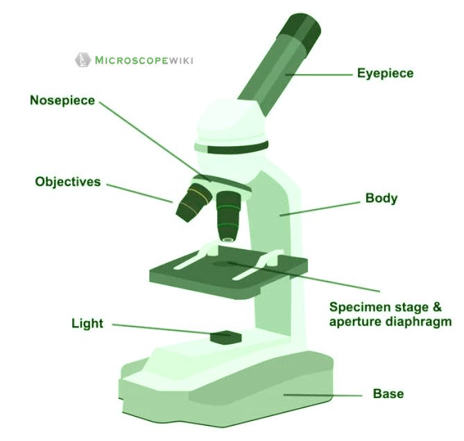
Microscope With Labeled Parts and Functions - 24 Hours Of Biology Optical parts and the functions The optical parts of the microscope are used to view, enlarge, and produce an image from a sample placed on a slide. These parts include Eyepiece: Eyepiece also contains ocular lens. It enhance the image of the viewer. This part is used for checking through the microscope. Eyepiece is found at the upper part of it. PDF Label parts of the Microscope Label parts of the Microscope: . Created Date: 20150715115425Z Microscope labeling and functions Flashcards | Quizlet Microscope labeling and functions STUDY Flashcards Learn Write Spell Test PLAY Match Gravity Created by mveet Terms in this set (27) Separates the eyepiece lens from the objective lenses Body Tube Holds the low-power and high-power objective lenses; allows the lenses to rotate for viewing Revolving Nosepiece Magnifies about 4x Parts of a Microscope and Their Functions - Microbiology Note Function: The arm Supports the head or body tube and connects it to the base of the microscopes. 3. Base. The bottom portion of Microscopes on which the arm portion is standing. It holds all the essential components. Application: The Base portion provides support to the microscope. Optical parts of a microscope and their functions
The Microscope [Parts & Functions] – HowForKids
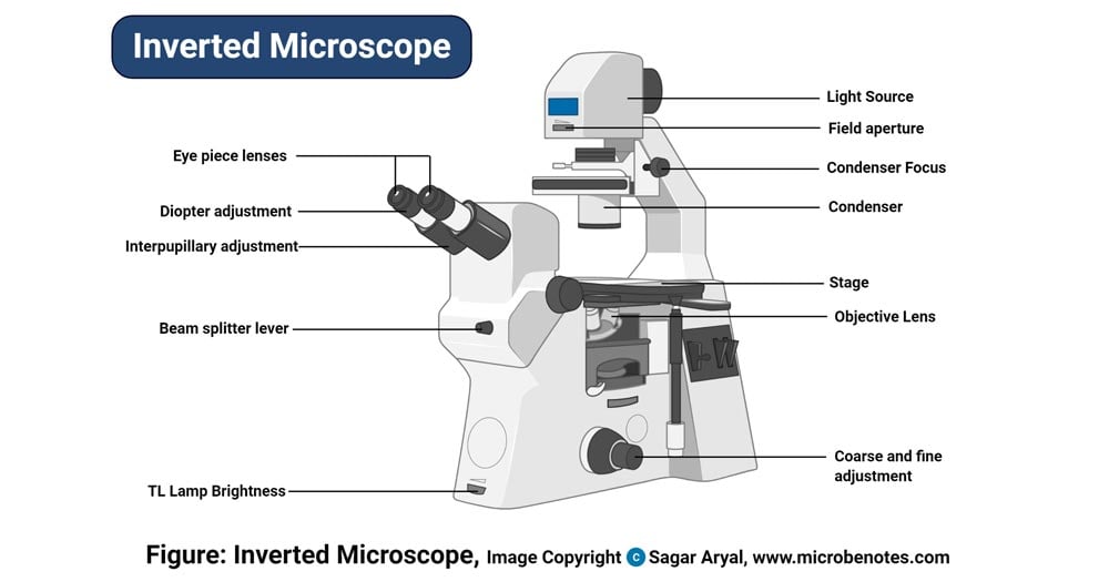
Binocular Microscope Anatomy - Parts and Functions with a Labeled ... Ocular lens or eyepiece of the microscope, Diopter adjustment of the eyepiece All of these parts are identified in a light microscope labeled diagram. So, first, make sure you can identify all these parts from this labeled diagram. Parts of the compound microscope
Understanding the Compound Microscope Parts and its Functions ...
Microscope Types (with labeled diagrams) and Functions Simple microscope labeled diagram Simple microscope functions It is used in industrial applications like: Watchmakers to assemble watches Cloth industry to count the number of threads or fibers in a cloth Jewelers to examine the finer parts of jewelry Miniature artists to examine and build their work Also used to inspect finer details on products
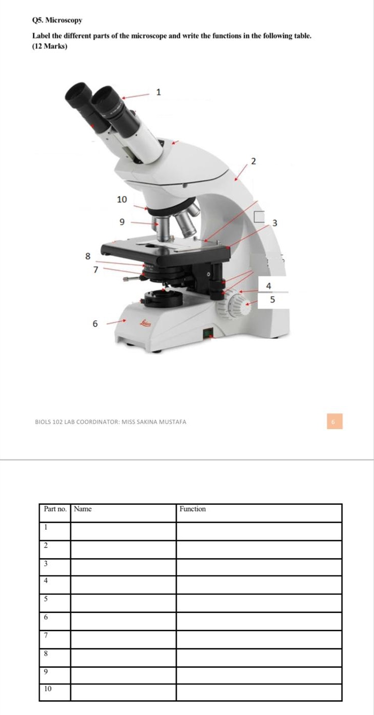
Solved Q5. Microscopy Label the different parts of the ...
(PDF) Introduction to Microscopy - ResearchGate 08.11.2017 · Functions of the Major Parts of a Optical Microscope [1-3] ... This new microscope doesn't require any special labels and could help increase access to low-cost plant science diagnostic tool.
Parts of a Microscope with Their Functions – Microbe Online
22 Parts Of a Microscope With Their Function And Labeled Diagram The field diaphragm control is located around the lens located in the base. Hinge Screw -This screw fixes the arm to the base and allow for the tilting of the arm. Stage Clips - They hold the slide firmly onto the stage. On/OFF Switch - This switch on the base of the microscope turns the illuminator off and on.
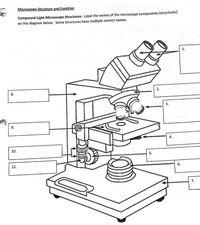
Answered: Microscope Structure and Function… | bartleby
Microscope Parts and Functions First, the purpose of a microscope is to magnify a small object or to magnify the fine details of a larger object in order to examine minute specimens that cannot be seen by the naked eye. Here are the important compound microscope parts... Eyepiece: The lens the viewer looks through to see the specimen.

The Microscope
Mastering Microbiology Ch 3 Flashcards | Quizlet Study with Quizlet and memorize flashcards containing terms like When observing a specimen in a microscope you are told that the total magnification of the specimen is 630x assuming you are using a standard occular lens with a magnification of 10x what is the magnification of the objective lens?, This activity asks you to sort items according to the most appropriate method for …

Microscope kaise use karen | What is microscope | Microscope parts and functions
Parts of Stereo Microscope (Dissecting microscope) – labeled ... Stereo microscopes (also called Dissecting microscope) are branched out from other light microscopes for the application of viewing "3D" objects. These include substantial specimens, such as insects, feathers, leaves, rocks, sand grains, gems, coins, and stamps, etc. Functionally, a stereo microscope is like a powerful magnifying glass.

Compound Microscope Parts, Functions, and Labeled Diagram ...
5 Types of Microscopes with Definitions, Principle, Uses, Labeled Diagrams 5 Types of Microscopes with Definitions, Principle, Uses, Labeled Diagrams March 1, 2022 by Sagar Aryal 5 Types of Microscopes Bright-Field or Light Microscope Dark Field Microscope Phase Contrast Microscope Fluorescence Microscope Electron Microscope Principle of Transmission Electron Microscope (TEM) References for types of microscopes

microscope | Types, Parts, History, Diagram, & Facts | Britannica
Light Microscope: Functions, Parts and How to Use It The function of the light microscope is based on its ability to focus a beam of light through a very small and transparent specimen, to produce an image. The image is then passed through one or two lenses for magnification to view. The transparency of the specimen allows for easy and fast light penetration. Specimens can vary from bacteria to ...

Microscope Parts and Functions
Parts of a Microscope Labeling Activity - Storyboard That Create a poster that labels the parts of a microscope and includes descriptions of what each part does. Click "Start Assignment". Use a landscape poster layout (large or small). Search for a diagram of a microscope. Using arrows and textables label each part of the microscope and describe its function.
microscope parts and functions - Quizizz
Electron microscope - Wikipedia An electron microscope is a microscope that uses a beam of accelerated electrons as a source of illumination. As the wavelength of an electron can be up to 100,000 times shorter than that of visible light photons , electron microscopes have a higher resolving power than light microscopes and can reveal the structure of smaller objects.
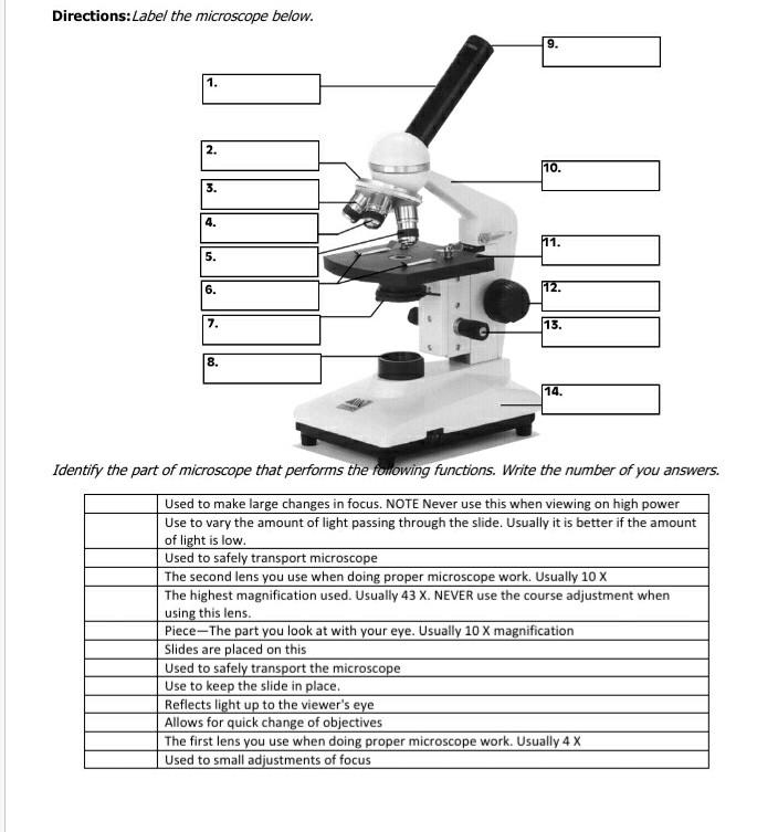
Solved Directions:Label the microscope below. 9. 1. 111 2 ...
Introduction to three-dimensional image processing skimage.exposure contains a number of functions for adjusting image contrast. These functions operate on pixel values. Generally, image dimensionality or pixel spacing does not need to be considered. Gamma correction, also known as Power Law Transform, brightens or darkens an image. The function \(O = I^\gamma\) is applied to each pixel in the ...

Microscope Parts and Functions Flashcards | Quizlet
Bright-field microscope (Compound light microscope) - Diagram (Parts ... Bright-field microscope parts (Labeled Diagram) Ocular Lens This microscope has two eye lenses or ocular lens on the top of the microscope that are used to focus the image from the objective lens. It is from these lenses that we see the magnified image of the specimen. Objective Lens

Microscope Parts & Functions - AmScope
Parts of the Microscope with Labeling (also Free Printouts) Let us take a look at the different parts of microscopes and their respective functions. 1. Eyepiece it is the topmost part of the microscope. Through the eyepiece, you can visualize the object being studied. Its magnification capacity ranges between 10 and 15 times. 2. Body tube/Head It is the structure that connects the eyepiece to the lenses.

Labeled Microscope Diagram | Microscope parts, Science fair ...
Introduction to three-dimensional image processing Introduction to three-dimensional image processing¶. Images are represented as numpy arrays. A single-channel, or grayscale, image is a 2D matrix of pixel intensities of shape (row, column).We can construct a 3D volume as a series of 2D planes, giving 3D images the shape (plane, row, column).Multichannel data adds a channel dimension in the final position …
Compound Microscope Parts, Functions, and Labeled Diagram ...
Microscope, Microscope Parts, Labeled Diagram, and Functions Illuminator: Illuminator is the most important microscope parts and it serve as light source for a microscope during slide specimen visualization. It is a continuous source of light (110 volts) used in place of a mirror. The mirror of microscope is used to reflect light from the external light source up through the bottom of the stage.
Parts of a microscope with functions and labeled diagram
Compound Microscope: Definition, Diagram, Parts, Uses, Working ... - BYJUS A compound microscope is defined as. A microscope with a high resolution and uses two sets of lenses providing a 2-dimensional image of the sample. The term compound refers to the usage of more than one lens in the microscope. Also, the compound microscope is one of the types of optical microscopes. The other type of optical microscope is a ...

Types, Parts and Functions of a Microscope
Nervous System – Medical Terminology for Healthcare Professions Introduction to the Nervous System. The picture you have in your mind of the nervous system probably includes the brain, the nervous tissue contained within the cranium, and the spinal cord, the extension of nervous tissue within the vertebral column.That suggests it is made of two organs—and you may not even think of the spinal cord as an organ—but the nervous system …

Lasec Education | Key parts of a compound microscope and how ...
A FINE GEORGE II MAHOGANY CASED CUFF PATTERN MONOCULAR MICROSCOPE A FINE GEORGE II MAHOGANY CASED CUFF PATTERN MONOCULAR MICROSCOPEJOHN CUFF, LONDON, MID 18th CENTURYThe body tube with stepped moulded shuttered eyepiece over ogee waist and objective tube incorporating marks for six positions on an exponential scale numbered 1 to 6, supported via a tapered collar set in a ring attached to a vertical slide moving against the fixed limb upright marked with six ...
22 Parts Of a Microscope With Their Function And Labeled ...
Microscope Quiz: How Much You Know About Microscope Parts And Functions ... This quiz will check how much do you know about Microscope Parts and Functions! The microscope has been used in science to understand elements, diseases, and cells. You must have used a microscope back in high school in the biology lab. Do you believe you understood how to use it? Take up the test and see. Questions and Answers 1. Arm: A.

Parts of the Microscope with Labeling (also Free Printouts ...
Microscope Parts and Functions Flashcards | Quizlet Magnifies image 40X found on the nosepiece. Base. Support/bottom of the microscope, used to carry the microscope. Light Source. Provides light to enable us to see the specimen on the slide. Arm. Used in order to carry the microscope. Coarse Adjustment. moves the stage up or down a lot, used first when viewing the slide.

Microscope Diagram Labeled, Unlabeled and Blank | Parts of a ...
Electron microscope - Wikipedia An electron microscope is a microscope that uses a beam of accelerated electrons as a source of illumination. As the wavelength of an electron can be up to 100,000 times shorter than that of visible light photons, electron microscopes have a higher resolving power than light microscopes and can reveal the structure of smaller objects.. Electron microscopes use shaped magnetic …

Compound Microscope Parts – Labeled Diagram and their ...
Label the microscope — Science Learning Hub Jun 08, 2018 · All microscopes share features in common. In this interactive, you can label the different parts of a microscope. Use this with the Microscope parts activity to help students identify and label the main parts of a microscope and then describe their functions. Drag and drop the text labels onto the microscope diagram. If you want to redo an ...
16 Types of Microscopes with Parts, Functions, Diagrams
Histology at SIU, tissue prep - Southern Illinois University ... Jun 16, 2022 · Modern cell biology uses many tools to reveal cell structures and functions that are not apparent on such routinely prepared slides. Many of these involve sophisticated reagents based on the specificity of enzymes, immunological antibodies, or gene sequences to label and localize specific proteins or other molecules.

Pin on Micellaneous
Microscope Parts & Functions - AmScope Microscope Parts and Functions Invented by a Dutch spectacle maker in the late 16th century, compound light microscopes use two sets of lenses to magnify images for study and observation. The first set of lenses are the oculars, or eyepieces, that the viewer looks into; the second set of lenses are the objectives, which are closest to the specimen.
Microscope parts and functions Quiz
Microscope World | Shop Microscopes For Every Application Microscope World | Shop Microscopes For Every Application

Compound Microscope Parts
A FINE GEORGE II MAHOGANY CASED CUFF PATTERN MONOCULAR MICROSCOPE A FINE GEORGE II MAHOGANY CASED CUFF PATTERN MONOCULAR MICROSCOPEJOHN CUFF, LONDON, MID 18th CENTURYThe body tube with stepped moulded shuttered eyepiece over ogee waist and objective tube incorporating marks for six positions on an exponential scale numbered 1 to 6, supported via a tapered collar set in a ring attached to a vertical slide moving …

Parts of a microscope with functions and labeled diagram
Parts of a Compound Microscope and Their Functions - NotesHippo Compound microscope uses in forensic labs it easy to detect human fingerprints. A compound microscope can be used to detect the presence of metals. The use of a compound microscope makes studying germs and viruses much easier. Compound microscope uses in schools makes learning biology easy for all children.

Microscope Diagram Labeled, Unlabeled and Blank | Parts of a ...
Label the microscope — Science Learning Hub 08.06.2018 · All microscopes share features in common. In this interactive, you can label the different parts of a microscope. Use this with the Microscope parts activity to help students identify and label the main parts of a microscope and then describe their functions.. Drag and drop the text labels onto the microscope diagram. If you want to redo an answer, click on the …

Microscope labeling and functions Flashcards | Quizlet

Microscope I Parts and function of Microscope I Scientech Biology I How to use microscope
Solved 5. Label the parts of the light microscope below ...

Compound Microscope Parts, Function, & Diagram | What is a ...
Microscope Types (with labeled diagrams) and Functions

Parts of a Compound Microscope and Their Functions

Solved tration Questions: (10 points) Label the diagram of a ...
Parts of Stereo Microscope (Dissecting microscope) – labeled ...

Biology Microscope Parts and Functions Diagram | Quizlet
Post a Comment for "38 microscope with labels and functions"