42 knee joint with labels
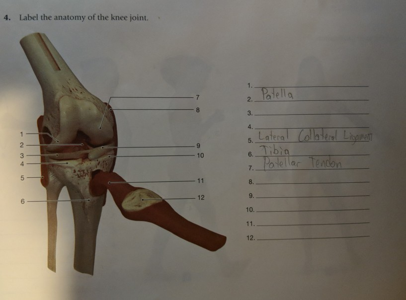
Label The Structures Of The Knee Joint - Solved Procedure 1 Identifying ... The knee joint is essentially made up of three bones: Start studying knee joint label. The femur (thigh bone), tibia (shin bone), and patella (kneecap) make up the bones of the knee. The medial and lateral menisci are fibrocartilage structures in the knee that serve two functions: The knee joint has three parts. The knee joint keeps these bones ... Label The Structures Of The Knee. Chegg - Solved Match The Knee Joint ... Structure of the knee joint 1. Ty label the structures of the knee joint (superior view by clicking and dragging the labels to the correct location lateral menis eu synovial . Label the structures of the knee. Label the structures of the knee. Tibia patellar surface lateral condyle of femur 16 medial condyle of femur anterior cruciate .
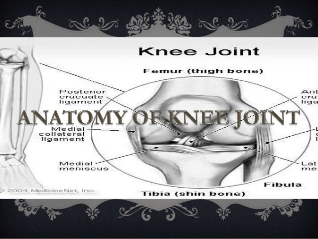
Knee joint: anatomy, ligaments and movements | Kenhub The tibiofemoral joint Medial condyle of femur Condylus medialis femoris 1/7 The tibiofemoral joint is an articulation between the lateral and medial condyles of the distal end of the femur and the tibial plateaus, both of which are covered by a thick layer of hyaline cartilage .

Knee joint with labels
A Diagrammatic Explanation of the Parts of the Human Knee Knee actually consists of three bones - femur, tibia and patella. Femur is the thigh bone, tibia is the shin bone and patella is the small cap like structure which rests on the other two bones. Femur is considered as the largest bone in the human body. The femur and the tibia meets at the tibiofemoral joint and patella rests on top of this joint. Knee Joint Picture Image on MedicineNet.com The knee functions to allow movement of the leg and is critical to normal walking. The knee flexes normally to a maximum of 135 degrees and extends to 0 degrees. The bursae, or fluid-filled sacs, serve as gliding surfaces for the tendons to reduce the force of friction as these tendons move. The knee is a weight-bearing joint. nurseslabs.com › 5-total-joint-knee-hip5 Total Joint (Knee, Hip) Replacement Nursing Care Plans Mar 18, 2022 · Joint replacements are indicated for irreversibly damaged joints with loss of function and unremitting pain, selected fractures, joint instability and congenital hip disorders. Total Joint Replacement can be performed on any joint except the spine. Hip and knee replacements are the most common procedures.
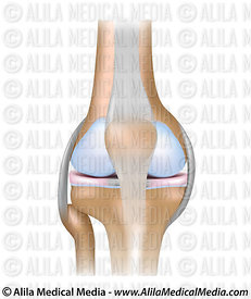
Knee joint with labels. Alila Medical Media | Knee joint, basic labels | Medical illustration Image size: 39.1 Mpixels (112 MB uncompressed) - 6250x6250 pixels (20.8x20.8 in / 52.9x52.9 cm at 300 ppi) A Labeled Diagram of the Knee With an Insight into Its Working Labeled Diagram of the Knee Joint Knee joint is one of the most important hinge joints of our body. Its complexity and its efficiency is the best example of God's creation. The anatomy of the knee consists of bones, muscles, nerves, cartilages, tendons and ligaments. All these parts combine and work together. shoulder joint with labels - Scottsdale Joint Center The Scottsdale Joint Center is in Arizona - Call us at 480-994-1149. Dr. Stuart Kozinn is an orthopedic surgeon in private practice in Scottsdale. Knee x-ray - labeling questions | Radiology Case | Radiopaedia.org Normal X-ray Knee - Frontal (with labels) Annotated image Frontal Knee Frontal 1. Femoral shaft 2. Patella 3. Base of patella 4. Apex of patella 5. Adductor tubercle of femur 6. Medial epicondyle of femur 7. Medial condyle of femur 8. Lateral epicondyle of femur 9. Lateral condyle of femur 10. Groove for popliteus 11. Intercondylar fossa 12.
Knee (Human Anatomy): Function, Parts, Conditions, Treatments - WebMD The knee is one of the largest and most complex joints in the body. The knee joins the thigh bone (femur) to the shin bone (tibia). The smaller bone that runs alongside the tibia (fibula) and the... Knee Joint Label Diagram | Quizlet Start studying Knee Joint Label. Learn vocabulary, terms, and more with flashcards, games, and other study tools. Label The Structures Of The Knee. - New Philippines expressways being ... Label the structures of the knee. To deepen the articular surface of the tibia, . The posterior and anterior cruciate ligaments (pcl and acl) limit forward motion of the knee bones, keeping them stable. The 3b scientific® anatomy video knee joint demonstrates the structure of the knee joint. Knee Joint - Anatomy Pictures and Information - Innerbody The knee, also known as the tibiofemoral joint, is a synovial hinge joint formed between three bones: the femur, tibia, and patella. Two rounded, convex processes (known as condyles) on the distal end of the femur meet two rounded, concave condyles at the proximal end of the tibia. Continue Scrolling To Read More Below... Additional Resources
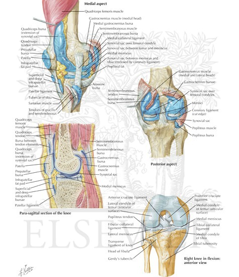
The Knee Joint - Articulations - Movements - TeachMeAnatomy The knee joint is a hinge type synovial joint, which mainly allows for flexion and extension (and a small degree of medial and lateral rotation). It is formed by articulations between the patella, femur and tibia. In this article, we shall examine the anatomy of the knee joint - its articulating surfaces, ligaments and neurovascular supply. Knee Joint - label pictures Flashcards | Quizlet Knee Joint - label pictures STUDY Flashcards Learn Write Spell Test PLAY Match Gravity Created by cfreynolds2018 Terms in this set (7) 1. Femur 2. Articular capsule 3. PCL 4. Lateral Meniscus 5. ACL 6. Tibia 1-6 7. Quadracep tendon 8. Suprapatellar bursa 9. Patella 10. Subcutaneous prepatellar bursa 11. Synovial cavity 12. Lateral Meniscus 13. Synovial Joint Diagram Label - schematron.org Start studying label the synovial joint. Learn vocabulary, terms, and more with flashcards, games, and other study tools. Labeled Diagram of the Knee Joint Knee joint is one of the most important hinge joints of our body. Its complexity and its efficiency is the best example of God's creation. The synovial membrane lines the entire joint except ... nurseslabs.com › 5-total-joint-knee-hip5 Total Joint (Knee, Hip) Replacement Nursing Care Plans Mar 18, 2022 · Joint replacements are indicated for irreversibly damaged joints with loss of function and unremitting pain, selected fractures, joint instability and congenital hip disorders. Total Joint Replacement can be performed on any joint except the spine. Hip and knee replacements are the most common procedures.
Print A&P Chapter 8 Joints flashcards | Easy Notecards
Knee Joint Picture Image on MedicineNet.com The knee functions to allow movement of the leg and is critical to normal walking. The knee flexes normally to a maximum of 135 degrees and extends to 0 degrees. The bursae, or fluid-filled sacs, serve as gliding surfaces for the tendons to reduce the force of friction as these tendons move. The knee is a weight-bearing joint.
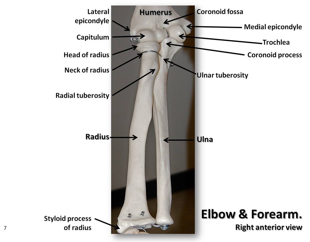
Bones of the elbow and forearm, anterior view with labels … | Flickr
A Diagrammatic Explanation of the Parts of the Human Knee Knee actually consists of three bones - femur, tibia and patella. Femur is the thigh bone, tibia is the shin bone and patella is the small cap like structure which rests on the other two bones. Femur is considered as the largest bone in the human body. The femur and the tibia meets at the tibiofemoral joint and patella rests on top of this joint.
Knee joint

Human Anatomy Lab: Knee Joint Model

Human Anatomy Lab: Knee Joint Model

Osteoarthritis of knee joint Poster | Zazzle.com
34 Label Knee Joint - Labels Information List

Health Leads UK Ltd: Supplements for Joint Health
KNEE JOINT

Alila Medical Media | Knee joint labeled drawing. | Medical illustration

Total Knee Replacement of the Right Knee | Total knee replacement, Knee replacement, Knee ...

Blog not found

Alila Medical Media | Knee joint, basic labels | Medical illustration

Medical suggestions
Alila Medical Media | Knee joint labeled. | Medical illustration
Welcome To Netter Images
Post a Comment for "42 knee joint with labels"